- STUDY OF THE MORPHOLOGY AND PHYSICO-CHEMICAL DISTRIBUTION (INCLUDING CRYSTALLINE COMPOSITION) IN MATERIALS AT THE MICRO AND NANOMETER SCALE
- ►Environmental scanning electron microscopy (ESEM)
- TEM
- AFM
- X-ray diffraction
- FETEM
- CATSUD-URV nanotechnology platform
- Confocal microscopy
- Z-sizer
-
Description
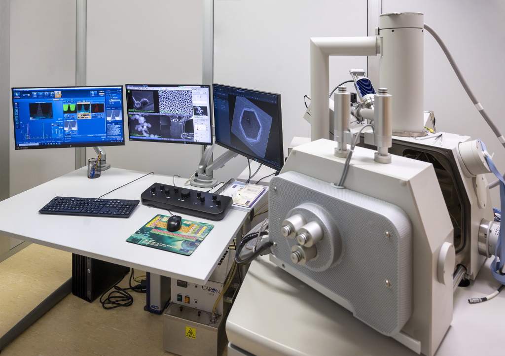
This microscope, the Quanta 600 from FEI company, uses an electron beam to form the image. It is characterized by its high resolution (3 nm) and its great depth of field that allows the focusing of different heights of the sample at the same time. Due to the electron-matter interaction, different signals that lead to different results are produced: topographic image, atomic number image contrast, energy dispersive X-ray spectroscopy, etc.
Conductive, non-conductive and incompatible with high vacuum specimens can be study using the three pressure modes that range from 10-5 mbar to 26 mbar (2600 Pa) with a resolution of 3nm. Humidity studies can be performed together with the Peltier stage from -20 °C a 50 °C. The microscope also has an oven that reaches temperatures up to 1500 °C, which allows to make temperature studies with the possibility to connect several gases. TEM grids which require to maintain the sample humidity can be observed with the Wet-STEM detector without the necessity of fixation, dehydration and inclusion.
Types of measurements achievable with the ESEM:
- Morphological and topographical observation of all type of specimens at 3 nm resolution
- Composition images (atomic number contrast)
- Energy dispersive X-ray spectroscopy (from boron). The elements present in the different parts of the sample can be obtained in a volume as small as a cubic micrometre
- Semi-quantitative analysis with 2% error. Element distribution maps
- Study of materials under heating and cooling. Dynamic experiments such as humidity cycles, temperature experiments (from -20 to 1.500 °C), etc.
- Observation in Wet-STEM mode
- Absorbed and induced current images (electron-beam induced current, EBIC)
-
Applications
- Particle analysis
- Morphological studies on food
- Contaminants
- Corrosion processes
- Coatings, metallurgic structures and ceramics characterization
- Pictorial layers
- Microelectronics
- Forensic applications
- Quality control
- Biomedical applications: cell cultures, tissues and organs study; etc.

-
Contact those
responsible - Mariana Stefanova Stankova, Ph.D.
- 977558123
- mariana.stefanova(ELIMINAR)@urv.cat
-
- Mercè Moncusí Mercadé
- 977558123
- merce.moncusi(ELIMINAR)@urv.cat
*Already registered on the User Portal?

