-
Description
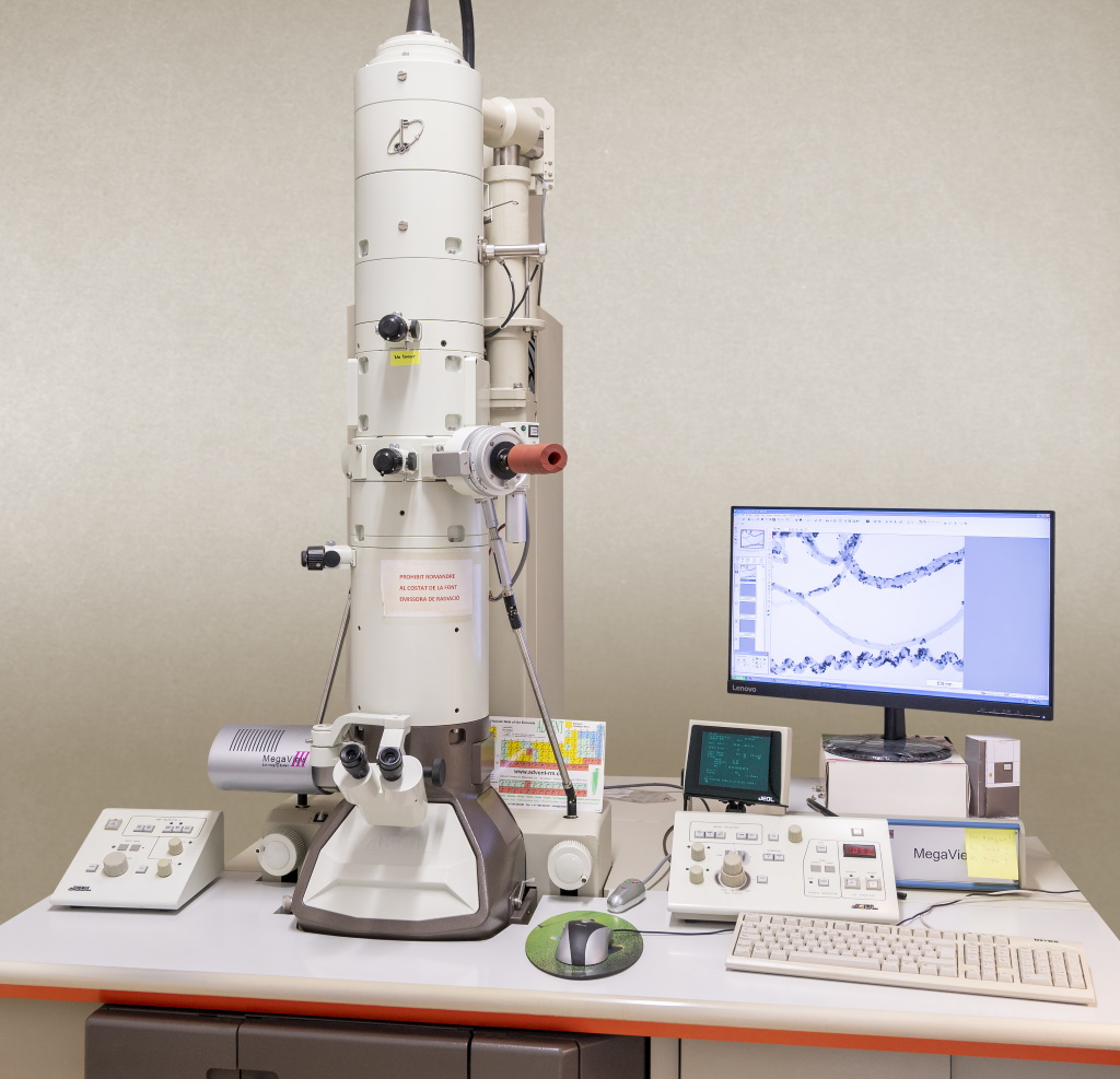
The JEOL 1011 is a high performance and high contrast TEM operated at 100 kV that is easy to use and projects excellent images. The TEM uses as a light source an electron beam generated by a tungsten filament. These electrons are accelerated and directed towards the sample by means of electromagnetic lenses in conditions of high vacuum. Electrons passing through the sample hit a phosphor screen that emits fluorescence.
The 0.2 nm resolution of this instrument allows the morphological study of particles and nanoparticles as well as their distribution. Thanks to ultramicrotomy or to the lamellas obtained by a FIB (Focused Ion Beam), it is suitable for biological applications, polymers and materials science
In life sciences, TEM allows the study of cytochemistry and immunocytochemistry at the cellular and subcellular scale, biological samples, such as animal and plant tissues, cell and bacterial cultures, viruses, subcellular structures, and macromolecules. In order to observe the sample under an electron microscope, it has to be prepared in advance according to specific methods of fixation, inclusion and ultramicrotomy.
The obtained image is flat, monochromatic (black and white) with magnifications ranging between 200x and 1.000.000x.
- Nanostructured material analysis (core-shell particles, thin films, nanorods, graphene, catalysers, etc.).
- Determination of morphology: shape, dimension and position of microcrystals or particles.
- Electron diffraction.
- Ultra-structural analysis of biologic samples.
- Cellular localization and colocalization of proteins using immunolocalization techniques.
- Location and distribution of enzymatic activities.
- Detection and distribution of specific carbohydrate residues and cytochemical studies
-
Contact those
responsible - Rita Marimon Picó, Ph. D.
- 977558473
- rita.marimon(ELIMINAR)@urv.cat
-
- Mercè Moncusí Mercadé
- 977558123
- merce.moncusi(ELIMINAR)@urv.cat
-
Technical Coordinator
- Mercè Moncusí Mercadé
- 977558123
- merce.moncusi(ELIMINAR)@urv.cat
*Already registered on the User Portal?