-
Description
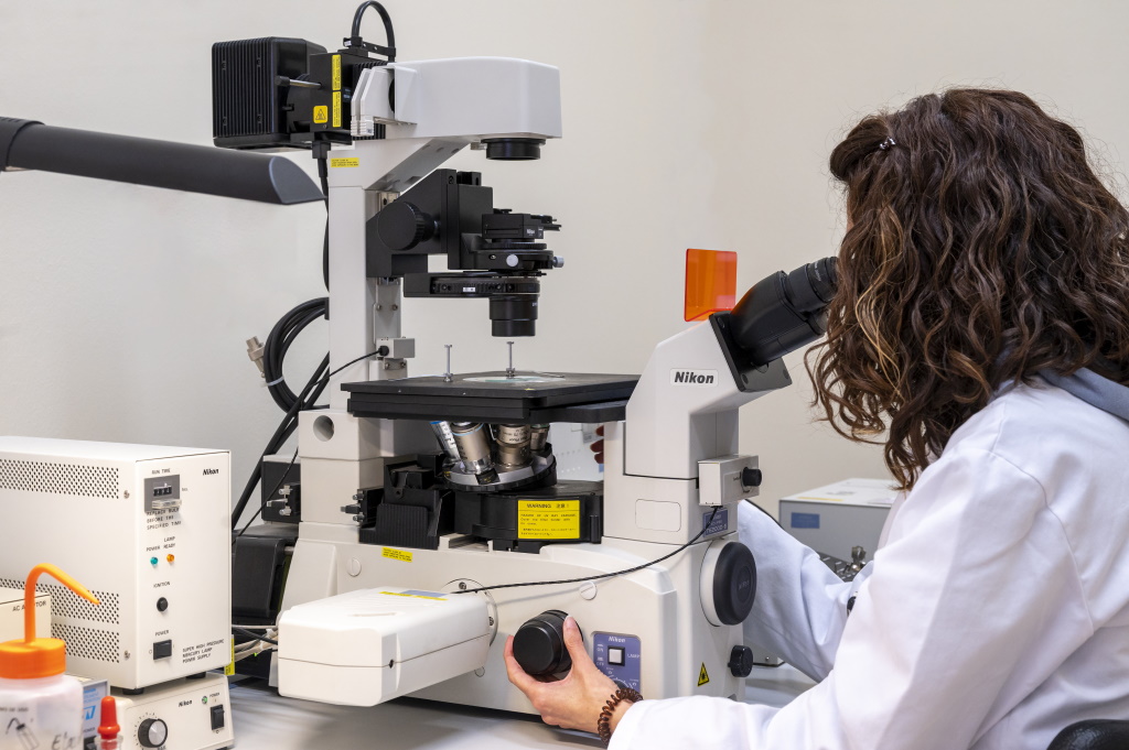
The Nikon TE 2000E confocal scanning laser microscope is an inverted optical microscope able to provide three-dimensional images of an object by gathering its fluorescence signal. It allows to eliminate the fluorescent signal coming from the sample that is out of focus, which is achieved in a non-destructive way, and thus provides optical sections from different planes of the sample. These sections can be used to make a three-dimensional analysis or to observe an internal part of the sample.
Confocal microscopy significantly increases the optical resolution and image contrast. These advantages make it an essential tool when it comes to obtain high-resolution images and when colocalization studies are carried out.
The optical sections obtained by confocal microscopy contain the information to be able to make a three-dimensional analysis: volumetric quantification, fluorescence distribution, colocalization analysis, etc.
The microscope is equipped with three excitation lasers (488nm, 543nm and 633 nm,) and four simultaneous detection channels (three fluorescence channels and one for transmitted light).
The HBO100 mercury lamp is used as a light source to visualize fluorescent samples; and the transmitted light for bright field and for differential interference contrast (DIC)
To view the sample, this system is equipped with the following filters:
- UV- Excitation BP 340-380; emission LP 425) for Alexa 405 o DAPI, etc.
- Blue- Excitation BP 450-490; emission LP 515) for FITC, Cy2, etc.
- Green- Excitation BP 515-560; emission LP 590) for TRITC, Cy3, etc.
- Red Excitation 633; emission 657/700) for Alexa 633, etc.
The objectives available in this microscope are:
- HC PL APO CS 10X/0.4 DRY
- HCX PL APO 20X/0.7 IMM
- HCX PL APO 20X/0.7 IMM (for water or oil)
- HCX PL APO CS 40X/1.25 OIL
- HCX PL APO 60X/ 1.4 OIL
- HCX PL APO CS 100X/1.4 OIL
- Three-dimensional analysis on biological samples: immunofluorescence in cells, tissues, biofilms, etc.
- Three-dimensional analysis of the colocalization of up to three markers.
- Studies of movement (FRAP) or interaction (FRET) of molecules in tissues and cells.
- Living cell studies: time lapse, intracellular ion measurement, 4D studies.
- Materials science: surface analysis, profilometry, roughness parameters, relief maps.
- In vivo and real-time cell analysis using fluorescent markers and/or fusion fluorescent proteins (GPF) and derivatives.
- Colocalization, internalization and intracellular transit studies.
- Physiological analysis of Ca2 + response.
- Do not put your hand nor expose your eyes through the optical path of the lasers.
- Put your sample before connecting the lasers.
- Do not directly observe the emission of the laser that hits the sample.
-
Contact those
responsibles - Dra. Rita Marimon Picó, Ph.D.
- 977558473
- rita.marimon(ELIMINAR)@urv.cat
-
- Mercè Moncusí Mercadé
- 977558123
- merce.moncusi(ELIMINAR)@urv.cat
-
Technical coordinator
- Mercè Moncusí Mercadé
- 977558123
- merce.moncusi(ELIMINAR)@urv.cat
*Already registered on the User portal?