-
Description
X-ray diffraction is a non-destructive technique to identify and quantify the crystalline phases present in solid matter. X-rays diffract into crystalline materials and produce characteristic diffractograms that act as fingerprints of each crystalline phase. The diffractogram reflects the crystal structure of the material (ordered arrangement of atoms in 3D space). The diffractogram is interpreted with the help of a database.
The technique is used to obtain information on the structure, composition and condition of polycrystalline materials. In addition to the identification and quantification of crystalline phases, it is also used in studies with variable temperature, measurements of cell parameters, degree of crystallinity , calculation of residual stresses, mosaicity , texture studies and adjustment of structures.
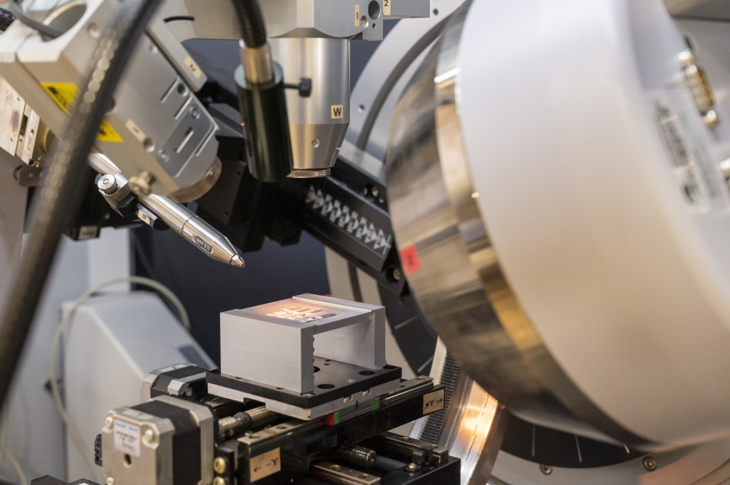
Bruker D8-DISCOVER
- VÅNTEC-500 area detector positionable from 90 to 300 mm of the sample
- 2D images of 2048x2048 pixels
- Göbel mirror for parallel optics
- He camera for the low angle area detector
- Video / laser system for positioning the sample
- ¼ Eulerian circle (0≤χ≤110 °, 0≤φ≤360 °) motorized in XYZ
- Collimators for microdiffraction of 50, 100 and 500 μm
- MRI chamber (-192 to 1300 °) with vacuum or non-reacting gas system
- Scintillation detector
- HUBER sample holder for rotating capillaries
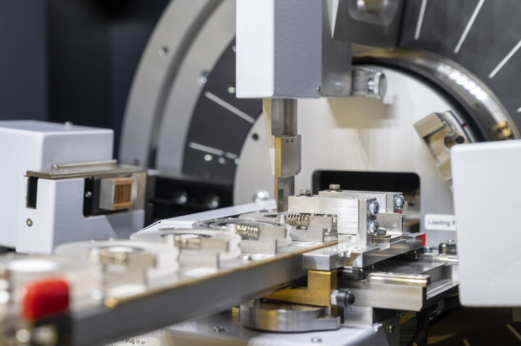
Bruker D8-ADVANCE
- 9-sample automatic loader with rotation / transmission
- Motorized divergent / parallel optics
- 1D LYNXEYE-XE-T detector
- MDT temperature chamber (-192 to 1300 °) with spinner rotation, motorized in Z, with vacuum system or non-reacting gas
- Vacuum secondary optics for measurements in SAXS mode
- HUBER sample holder for rotating capillaries (Debye-Scherrer geometry)
- Si(510) low background sampleholders
- Tighed sampleholders for sensitive samples
- Sample holder for transmission (Bragg- Brentano geometry)
Software
- TOPAS 6.0
- Diffrac.Suite 5.2
- PDF-2 2018
- Multex Area 3.0
- GADDS
- LEPTOS 7.0
- Diffrac.SAXS
- Diffrac.TEXTURE
- Qualitative / quantitative analysis of crystalline phases (Rietveld method).
- Cluster Analysis.
- Quantification of amorphous and degree of crystallinity.
- Crystallite size (crystallite size) and microdeformation.
- Stability of crystalline phases with temperature.
- Determination of thermal expansion coefficients.
- Transformation kinetics study.
- Measurement of residual stress.
- Determinació de l'orientació de monocristalls.
- Analysis of the texture of a material.
- Determination of the orientation of single crystals.
- Rocking - curve studies.
- Microdifraction (up to 50 microns).
- Obtaining Laue photographs.
- Qualitative analysis of thin films.
- Determination (indexing) and adjustment of reticular parameters.
- Determination of the thickness of thin films (XRR).
- Analysis of thin films by grazing incidence (GIXRD).
- Low angle analysis (> 0.5 ° 2θ) in 1 and 2D (SAXS).
- Organic and inorganic solid samples (powder or bulk)
- Membranes & filters
- Capillaries and fibers
- Irregular archaeological samples (max 80x80x40mm)
- Thin Films
- Electrical circuits
- Environmentally sensitive samples Monocristalls
- Single crystals

-
Contact the person
in charge - Dr. Francesc Gispert i Guirado
- 977559783
- drx.srcit(ELIMINAR)@urv.cat
-
Technical coordinator
- Ramon Guerrero Grueso
- 977558149
- rmn.srcit(ELIMINAR)@urv.cat
*Already registered on the User Portal?
The transfer of knowledge:
DIFFRAC.EVA and TOPAS training courses


